Olympus is continuing to search for the best light microscopy images in 2021. Olympus’ third Global Image of the Year Life Science Light Microscopy Award recognizes the very best in life science imaging worldwide. Participants can win an SZX7 microscope with a DP28 digital camera or a CX23 microscope.
One global winner and one regional winner from the participants in each of the three regions (Asia-Pacific, Europe, the Middle East and Africa, and Americas) will be selected.
Global prizeSZX7 Stereo Microscope with DP28 Digital CameraSZX7 Stereo Microscope
Click here to read more about the SZX7 microscope DP28 Digital Camera
| Regional prize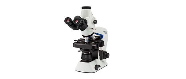 CX23 Upright Microscope
|
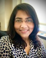
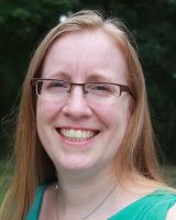 | Wendy Salmon, Director, Hooker Imaging Facility in the Cell Biology at the University of North Carolina at Chapel Hill School of MedicineWendy Salmon (she/her/hers) is Director of the Hooker Imaging Facility in the Cell Biology at the University of North Carolina (UNC) at Chapel Hill School of Medicine as of September 2021 and co-directs the annual Analytical and Quantitative Light Microscopy short course at the Marine Biological Laboratory (MBL) in Woods Hole, MA, USA. She studied biology at the University of Richmond in Richmond, VA, USA and discovered the wonders of light microscopy during summer research at the UNC in her hometown of Chapel Hill NC, USA. She received early training in live cell imaging with Dr. Clare Waterman and Dr. David McClay before transitioning to core facility work in 2002. She has advised and trained hundreds of researchers in basic and advanced light microscopy at four institutions and the MBL, most recently at the Whitehead Institute in Cambridge, MA, USA. She previously served as a judge for the Olympus BioScapes and Koch Center Image Awards contests. |
 | Geoff Williams, Manager of the Leduc BioImaging Facility at Brown UniversityGeoff Williams is in his fourteenth year as manager of the Leduc BioImaging Facility at Brown University. The opportunity to combine visual arts, science, technology and mastery of a skill clicked with his discovery of Microscopy (electron and light) as an undergraduate at Connecticut College. Geoff transitioned from a graduate program at Michigan State University to running the Imaging facility at Central Michigan University before arriving at Brown. Over the past 20 plus years he has been honing his craft as both an electron and light microscopist, paying much more attention to the aesthetic of each image collected than is typically required of a purely scientific investigation. Geoff’s work, under the name Nanoscape, provides a tactile and striking view of samples we may or may not encounter in our day-to-day lives. |
 | Harini Sreenivasappa, Manager of Cell Imaging Center at Drexel UniversityHarini Sreenivasappa is the Manager of Drexel University’s light microscopy core facility, the Cell Imaging Center. She was introduced to microscopy during graduate school at Texas A&M University where she studied the role of microenvironment stimuli on cellular sensing and adapting as it takes place in blood vessel wall remodeling in cardiovascular disease. This eventually led to a PhD in Biomedical Engineering. She has over 10 years of experience working with various microscopy techniques such as Atomic Force Microscopy (AFM), spinning disk confocal, and Total Internal Reflection Fluorescence (TIRF) microscopy. With ASCB’s COMPASS Outreach grant, she created and curated a Traveling Micrographs exhibit showcasing micrographs by Texas A&M University’s (TAMU) researchers that was free and open to the public. The goal of the series of exhibits was to share research at TAMU with the local community and stimulate interest in imaging science. |
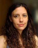 | Siân Culley, Postdoctoral Research Associate, MRC-LMCB, UCLSiân Culley first started using microscopes in a summer research project in 2009 studying calcium signaling in the mouse cochlea and has been imaging ever since. She did her PhD at University College London, where she developed a novel STED microscopy technique and investigated the underlying photophysics of CW-STED. Since 2014 she has worked as a postdoc at the MRC Laboratory for Molecular Cell Biology, UCL, with Professor Ricardo Henriques. Siân is currently developing novel methods for super-resolution microscopy, particularly through open-source image analysis. She also enjoys disseminating these techniques through teaching on courses and conferences, and in conjunction with the Royal Microscopical Society established the "Women in Microscopy" online resource. |
 | Stefan Terjung, Operational Manager of the ALMF at EMBL HeidelbergStefan Terjung studied biology and chemistry at the University of Heidelberg (DE). At the beginning of his studies he discovered his passion for microscopy techniques. For his thesis at the Institute of Cell Biology he investigated biological applications of two-photon microscopy. He obtained his PhD in botany at the Heidelberg Institute for Plant Sciences (HIP) in 2004. Stefan joined the Advanced Light Microscopy Facility (ALMF) at EMBL Heidelberg in 2003. Since 2016 he is Operational Manager of the ALMF. In this position he is regularly involved in organizing and teaching courses on light microscopy techniques. |
 | Rachid Rezgui, Research Instrumentation Scientist Microscopy, New York University Abu DhabiRachid studied Physics at the Leibniz University of Hanover in Germany and did his PhD in Biophysics at the Ecole Polytechnique in France studying DNA-Protein interactions at the single molecule level. He joined the microscopy core facility at New York University Abu Dhabi in 2014 and has since been involved with all types of microscopes (Two-Photon, Super-Resolution, Confocal, Fluorescence Lifetime, Widefield, etc.). Rachid is a microscopist and an active research scientist. He is involved in all aspects of optical imaging like sample preparation, training, acquisition and post-processing as well as core facility management. |
 | Wen-Biao Gan, Director of Institute of Neurological and Psychiatric Disorders at Shenzhen Bay LaboratoryWen-Biao Gan obtained his Bachelor’s degree in laser physics at Tsinghua University in 1986, and Ph.D. degree in neurobiology at Columbia University in 1995. He was a tenured professor in neuroscience at New York University School of Medicine in 2012. He has done a series of studies in the fields of learning and memory, sleep functions and microglia biology in the past 20 years. |
 | Anne Beghin, Assistant Professor, Research, Mechanobiology Institute, National University of SingaporeDr. Anne Beghin is a multidisciplinary scientist with fifteen years of extensive research experience across academia and industry. She obtained her PhD in oncology in 2007 at the University Claude Bernard in Lyon (France). She then moved to optical microscopy at the Université de Lyon, where she established the microscopy platform and developed live cell imaging solutions and image analysis services for 4 years. Subsequently, she moved to the Mechanobiology Institute (MBI) in Singapore to study organoids using advanced imaging and HCS. This work has resulted in a patent and publications are on-going. |
Download the Image of the Year Award 2020 wallpaper package now for free and beautify your screen!
Download the wallpaper package for desktop (ZIP, 40.2 MB)
Download the wallpaper package for mobile (ZIP, 26.8 MB)
Download your favorite virtual backgrounds and add them to your meetings!
Download the virtual backgrounds package for your meeting application (ZIP, 3.36 MB)