We are pleased to share the Phase 1 winners of the Complement-ARIE NAMs Reduction to Practice Challenge. You can learn about all of the teams participating below.
Predictive Brain Combinatorial NAM
Submitted by Massachusetts General Hospital
Team Captain: Alice Stanton
Team Members: Haroon Kalam, Venkatesh Pooladanda, Romane Gaston-Breton, Louis DeRidder
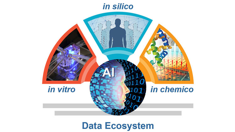
Our team is creating a new tool, Predictive Brain Combinatorial NAM, to help scientists predict how humans will respond to treatments and toxins. Traditional animal tests often fail to predict how treatments will work in people, especially for brain diseases like Alzheimer's or Parkinson's, leading to many failed drugs and delaying treatment options for the people who need them. Our approach combines three methods in a smart loop: computer simulations (using a powerful AI foundation model) to first predict effects, lab-grown mini-brains-on-chip (organoids) made from human cells to test and refine those predictions and quick chemical tests to guide the process. Over time, the AI gets better through iteration, so eventually, new compounds can be predicted just by the computer without always needing the mini-brains.
We have already built advanced mini-brains with all key cell types that demonstrate improvements over existing approaches, tested them with various chemicals, and used AI to analyze and improve results. This could make drug development faster, safer, and more accurate, helping protect people from harmful substances and advancing treatments for brain disorders. It is a highly innovative approach, filling gaps in current tools, scalable, focuses on real human biology, and builds toward a future where we identify beneficial treatments rapidly.
Combining vTIME and AI for IO Drug CRS Prediction
Submitted by Qureator Inc.
Team Captain: Jungfeng Wang
Team Members: Byungjun Lee, Aneesh Sathe, Sanghee Yoo
Immunotherapies like T-cell engagers are powerful cancer treatments, but risk triggering a deadly "immune storm" called Cytokine Release Syndrome (CRS). Currently, researchers test drugs on animals or in petri dishes to predict safety. However, animal biology differs fundamentally from humans, missing severe vascular toxicities. Conversely, petri dishes lack blood vessels and physical barriers, creating false alarms. Consequently, many promising drugs unexpectedly fail in human trials.
We propose a fully human, AI-driven alternative to animal testing. Using a "tumor-on-a-chip" technology called vTIME, we grow 3D human tumors connected to living human blood vessels and immune cells, accurately mimicking drug delivery and tissue interaction.
To analyze these complex reactions, we utilize QuriCore, an advanced AI. It evaluates 3D microscopic images to precisely measure cancer destruction. Simultaneously, it tracks blood vessel collapse and inflammatory protein surges to generate a highly accurate, multi-dimensional safety score.
By testing drugs with known toxic histories against safer profiles (like TGN1412 and MGD009), we will prove this platform precisely predicts human immune reactions. Having recently secured a historic FDA trial approval using vTIME data instead of animal testing, this technology will make clinical trials safer, accelerate cancer cures, and drastically reduce animal use.
Human Lung Models for Anti-Fibrotic Drug Testing
Submitted by Wake Forest University Health Sciences
Team Captain: Robyn Gore
Team Members: Sean V. Murphy, Steven R. Bauer, Sean Porazinski, Timothy Leach, Alexandria Pendino
Lung scarring, known as fibrosis, affects millions of people worldwide and has very few effective treatments. Researchers currently rely on animal testing to evaluate new drugs, but these models often fail to predict how drugs will work in humans, leading to repeated clinical trial failures.
Our project develops a two-part human cell-based testing system to replace animal models in anti-fibrotic drug evaluation. The first part is a high-speed automated platform that tests large numbers of drug candidates using miniature 3D printed human lung tissue models. The second is a more complex 3D lung tissue model that closely replicates human lung structure and function, allowing researchers to confirm that promising drugs work at the tissue level.
We will test drugs already known to have failed in human clinical trials to demonstrate that our system can identify ineffective treatments before they reach patients, something current animal models cannot reliably do. The system can also detect drug toxicity early in the testing process.
Our team includes university researchers with tissue modeling expertise, an industry partner that manufactures the 3D bioprinting platform, and a former senior FDA Branch Chief who will ensure the system meets regulatory standards. This platform could significantly reduce animal use in drug development while providing more human-relevant results, ultimately accelerating delivery of effective treatments to patients.
Combinatorial NAM for Human Atherosclerosis Model
Submitted by Endomimetics Inc.
Team Captain: Joseph Garner
Team Members: Jun Chen, Jennifer Sherwood, Ho-Wook Jun, Brigitta Brott, Kyungsang Kim
Heart disease is the leading cause of death worldwide, and atherosclerosis is its most common underlying cause. Many drugs that appear promising in early testing fail in human clinical trials because current models do not accurately reflect human disease. Animal studies are costly, slow, and often poorly predictive, while traditional laboratory tests rely on simplified cell cultures that lack the structure and biological complexity of human arteries. As a result, there is an urgent need for more advanced, human-relevant platforms. This project will develop AtheroVasc, a laboratory-grown human vascular model that closely mimics early atherosclerosis. Built using human cells, it recreates the natural three-layer structure of arteries and key early disease features.
Using this system, we will establish three complementary laboratory tests to measure immune cell buildup, fat accumulation, and inflammation, the processes that drive early plaque formation. These tests will run in standard multi-well plates, enabling efficient and scalable drug evaluation. AI-assisted analysis will automatically interpret results, improve consistency, and reduce bias. By combining realistic human biology, scalable testing, and automated analysis, this project provides a more predictive and ethical approach to accelerating early-stage atherosclerosis drug development while reducing reliance on animal testing.
CAR-T Immunotherapy Clinical Trials on a Chip
Submitted by CAR-T Chips Team
Team Captain: Weiqiang Chen
Team Members: David Baek, Lunan Liu, Tamas Gonda, Saba Ghassemi
Pancreatic cancer remains one of the most difficult cancers to treat. Even advanced immunotherapies like CAR-T cell therapy, which reprogram a patient's own immune cells, often fail against this disease. This failure is largely due to the heterogeneous and "immune cold" nature of pancreatic tumor. Currently, scientists test new therapies mostly in mice. But mice don't have human tumors and human immune systems, so results often don't translate to patients. Compounding this, no reliable clinical method exists to quickly and accurately predict which patients will benefit from CAR-T treatment. To address these challenges, our team developed an innovative solution combining three cutting-edge technologies, patient-derived organoids, organ-on-a-chip platforms, and artificial intelligence (AI) to predict patient responses to CAR-T therapy. Our approach uses a patient's own cells to build a miniature tumor replica on a microfluidic chip, then employs AI to analyze complex treatment processes and forecast therapeutic outcomes. This platform allows us to observe, in real time, whether CAR-T cells can find, infiltrate, and kill that specific patient's tumor, providing answers in weeks instead of months while reducing animal testing. By serving as a new clinical tool implementable directly in practice, our solution establishes a "clinical trials on a chip" paradigm that helps doctors choose the right treatment for the right patient—bringing us closer to truly personalized cancer care.
.
AI-integrated human skin immune organoids
Submitted by Stanford University
Team Captain: Calvin Kuo
Team Members: Barbara Engelhardt, Hudson Horn
Many promising medicines fail late in development because preclinical tests often do not predict what will happen in people. Animal studies and simplified cell models can miss key human biology, leading to drugs that do not work as expected or cause unanticipated side effects. This is especially important in skin, where drug benefit and drug rashes are driven by complex interactions between immune cells, support cells, and the skin barrier.
We will build a human focused testing system that utilizes a complex human tissue model called organoids that preserve multiple layers of human skin and different cell types and will combine this with a multimodal measurement system that shows that the tissue is doing (what cell types are activated and what they’re doing) and artificial intelligence that integrates these measurements into clear predictive readouts.
The outcome will be a “digital twin” of human skin responses that can predict whether a treatment will reduce inflammation, promote repair, or cause harm such as skin damage. We will also identify biomarkers that predict who is most likely to respond and who may be at higher risk of side effects, helping make clinical trials smaller, faster, and safer. Because it is trained and validated on human tissue, this platform can reduce reliance on animal testing and can be adapted beyond skin to other organs over time.
Combinatorial Human Bone Remodeling NAM Platform
Submitted by University of Massachusetts - Amherst
Team Captain: Jungwoo Lee
Team Members: Patrick Ryan, Leili Shahriyari, James Chambers, Jun-Goo Kwak
Osteoporosis is a common age-associated degenerative disease, often termed a silent killer because it remains undetected until life-threatening fractures occur, making it a major public health concern. Mature bone maintains strength via continuous formation and resorption, and osteoporosis results from chronic remodeling imbalance. Progress in treatment and prevention is limited by the difficulty of detecting subtle, long-term effects of drugs and environmental exposures using animal models and clinical studies, underscoring the need for New Approach Methodologies (NAMs) that reproduce human bone complexity and metabolism.
We developed lab-grown bone models that replicate the structure and cellular activity of mature human bone, including bone-forming and bone-resorbing cells on the surface and sensor cells embedded within. These models reproduce the natural cycle of bone breakdown and rebuilding under controlled conditions while enabling real-time imaging and longitudinal biochemical profiling. This quantitative platform supports computational modeling. By integrating tissue models (in vitro), biochemical measurements (in chemico), and computational simulations (in silico), we aim to establish a predictive human bone remodeling NAM platform. We will demonstrate its utility in two contexts: optimizing osteoporosis drug sequencing and screening environmental factors that impair bone health. This work has the potential to establish a new standard for bone remodeling NAMs.
PACE-IT: Immune-Cardiac NAM for Med Device Testing
Submitted by University of Minnesota
Team Captain: Brenda Ogle
Team Members: Andrew Khalil, Doug Hine, Maggie Pistella
Pacemakers are life-saving medical devices that help the heart beat normally. Before new pacemaker electrodes can be used in patients, they must be tested for safety and performance. Currently, these tests are done mostly in animals, which is expensive, slow, and not always accurate for predicting what happens in humans. Our team proposes to build a living human heart model in a dish that can be used to test pacemaker electrodes directly in human tissue and limits the use of animals. This “testbed” will be made from human stem cells (not embryonic) that are turned into beating heart cells and immune cells, forming a small piece of living heart tissue. Real pacemaker electrodes can then be attached to this tissue to study how well they work and how the tissue responds over time. This innovation will make pacemaker testing faster, cheaper, and more human-relevant. Instead of spending over $20,000 to test a single device in animals, our human testbed will cost approximately $500 per test. It will also provide valuable information about inflammation, healing, and electrical function to help scientists design safer, more effective devices. The project brings together experts from Medtronic, a leader in pacemaker technology, and the University of Minnesota, a global leader in heart tissue engineering. In the future, this technology could also be used to test other implanted devices and accelerate the discovery of better treatments for heart disease.
High-Throughput Human Tissue Remodeling & Toxicity
Submitted by First State Click-Chem Solutions
Team Captain: Lucas Lu
Team Members: Joseph Fox, Ilya Safro, Michael Axe, Zugui Zhang
We are developing a human-based testing platform to make drug development faster, safer, and less dependent on animal studies. Many diseases, including arthritis, organ scarring (fibrosis), and cancer, are driven by changes in the tissue “matrix,” the natural scaffolding that supports cells. Because animal matrix can differ from human matrix, drugs that look promising in animals may fail in people or cause unexpected side effects.
Our solution, the Molecular Highlighter, uses a Nobel Prize-recognized technique called click chemistry. We test tiny pieces of donated human tissue, smaller than a grain of rice, and label the newly made matrix as cells build it. This lets us measure, in real time, whether a drug improves tissue repair or triggers hidden damage, a functional readout that standard tests often miss.
We combine this assay with AI to prioritize which medicines to test and to explain why certain drugs are likely to work. We then compare our findings with long-term health records to confirm that the lab results align with real-world outcomes. This integrated approach can accelerate the discovery of safer treatments and may help doctors use a patient’s own biopsy to select the most effective chemotherapy dose.
Combinatorial NAM for Endometriosis Drug Efficacy
Submitted by VeriSIM Life, Inc.
Team Captain: Jyotika Varshney
Team Members: Morgan M. Stanton, Hugh S. Taylor, Divesh Bhatt, Roshan Bhave
Endometriosis affects roughly 1 in 10 women of reproductive age, yet available treatments remain limited. One reason is that animal models used early in drug development poorly predict what will work in patients. Mice and rats do not naturally develop endometriosis, and surgically created versions do not faithfully reflect human disease. Many promising compounds advance based on misleading animal results, only to fail in clinical trials.
Opal Therapeutics and VeriSIM Life are building a translational platform to reduce dependence on animal models and improve early decision making in endometriosis drug development.
First, Opal grows patient derived uterine organoids that replicate the cellular architecture and signaling environment of human tissue. These models allow drug candidates to be tested directly in human biology, capturing disease relevant responses that animal systems often miss. Second, VeriSIM Life integrates those laboratory readouts into its AI driven mechanistic modeling framework, translating molecular and phenotypic signals into quantitative predictions of clinical efficacy and safety.
By combining human tissue biology with predictive computational modeling, this approach enables more confident go or no go decisions before committing to costly and translationally unreliable animal studies. The platform could accelerate timelines, improve efficiency, and reduce unnecessary animal testing while advancing new therapies for women living with endometriosis.
Multi-Brain Region Assembloid Combinatorial Models
Submitted by Regenerative Research Foundation
Team Captain: Jeff Stern
Team Members: Sally Temple, Taylor Bertucci, Steven Lotz, Catherine Hamann, Thomas Kiehl, Kristina Roberts, Michelle Lewis
Stem cells have the remarkable ability to self-organize into small organs termed organoids that express characteristics of native brain composition. We will combine brain region-specific organoids into multiregional assembloids that express interactions critical for brain development, function and disease. The Neural Stem Cell Institute (NSCI) has developed protocols to more efficiently produce brain region-specific organoids. The ready availability of these organoids enables NSCI to take the next step- combining organoids from different brain regions to form assembloids that model the interregional interactions that commonly occur in the brain. Assembloids will be created by simply adding more than one type of organoid to a specialized culture dish where the organoids fuse to form assembloids. The response of the combinatorial assembloids will be used to evaluate the neurotoxicity of therapeutic candidates under development to treat neurologic and psychiatric disorders. The organoid and assembloid models will complement and reduce the use of animal models for drug development.
Multiorgan Predictive Biologic Toxicity Framework
Submitted by Biopico Systems Inc.
Team Captain: John Collins
Team Members: Henry Wong, Anshu Agrawal, Johar Kohana
Multi Organ Predictive Analysis with Computational Twin addresses a major challenge in developing biologic medicines. Some immune based drugs, including monoclonal antibodies and checkpoint inhibitors, can cause serious side effects when the immune system becomes overactive. These reactions are difficult to predict before clinical trials. Current safety testing relies heavily on animal studies, which are costly and may not accurately reflect how the human immune system responds. In addition, known differences between males and females in immune responses aren't always considered in early safety testing.
Our solution is a human based laboratory platform that combines engineered human tissues with circulating immune cells under controlled flow conditions. We measure immune signals and markers of tissue injury to see how the immune system affects organs. These experimental results are then connected to a computational framework that models dose response, individual variability, and sex related differences. Together, this integrated system translates laboratory data into clear risk categories that can help guide safer dose selection.
By directly modeling human immune and organ interactions, this approach aims to improve prediction of immune related toxicity, reduce reliance on animal testing, and support more personalized and reliable safety assessment of new biologic therapies.
Human Brain Organoids to Predict DNT
Submitted by Bioprinting Laboratories Inc.
Team Captain: Pranav Joshi
Team Members: Xuexia Wang, Lin Li, Minseong Lee
Neurodevelopmental disorders affect nearly one in six children in the United States and are a growing public health concern. Although genetics play important role, increasing research has shown that exposure to certain medications, drugs, alcohol, and environmental chemicals during pregnancy may raise the risk. Despite the developing brain being especially sensitive, only a small fraction of commonly used chemicals has been tested for their effects on brain development. Current testing relies largely on animal models, which are slow, expensive, and do not always reflect how the human brain responds. Our team is developing a faster, more human-relevant way to evaluate chemical safety using lab-grown human brain organoids - tiny, three-dimensional tissues that mimic early brain development. These organoids will be grown on a proprietary culture platform designed to improve nutrient and oxygen delivery and support realistic, long-term chemical exposure. We will examine how chemicals affect brain growth, cell health, neural communication, and gene activity, and use advanced data analysis to help predict potential developmental risks. Building on our prior research, this project aims to improve chemical safety testing, reduce reliance on animal research, and support better regulatory decisions. Ultimately, this work will enable early identification of harmful exposures to children and help protect them during critical periods of brain development.
Precision dosing of oligonucleotide therapeutics
Submitted by Javelin Biotech Inc.
Team Captain: Murat Cirit
Team Members: Shiny Rajan, Piet van der Graaf, Suruchi Bakshi, Emily Geishecker
Many therapies for rare diseases use liver-targeted oligonucleotides as genetic switches to silence harmful proteins or correct metabolic imbalances. However, development is often hindered by biological differences between humans and animals, causing dosing strategies validated in animals to lead to unexpected liver toxicity or treatment failure in patients. Historically, these failures have resulted in clinical trial terminations, safety warnings, and the withdrawal of life-saving drugs.
This project introduces a human-centric methodology to evaluate candidates before trials by integrating two technologies. First, a Liver-on-a-Chip uses human cells to create a functioning 3D model that mimics blood flow and organ function. This allows us to measure drug movement and detect long-term safety issues animal tests miss. Second, a Digital Twin platform integrates this data into computer models to generate virtual patient cohorts, simulating how individuals with various genetics and metabolism will respond.
We are qualifying this method using Fitusiran, a hemophilia drug. Early Fitusiran trials struggled because doses were too high for some patients, causing safety concerns. By using our liver-on-a-chip and digital twin system, we can accurately identify the appropriate dose for patients. This approach makes drug development faster, safer, and more efficient, ensuring the right dose reaches the right patient.
NeuroTraX-AI
Submitted by Frontier Bio Corporation
Team Captain: Eric Bennet
Team Members: Sam Pashneh-Tala, Julia Schachenhofer, Nigel Gomes
NeuroTraX-AI: A Combinatorial NAM for Human BBB Integrity
The blood-brain barrier (BBB) is a crucial protective layer that shields the brain from toxins, but it also creates a major hurdle because it blocks life-saving medicines from reaching brain tissue. Currently, scientists rely heavily on animal testing to study this barrier, but animal brains often react differently to drugs and injuries than human brains do.
NeuroTraX-AI solves this problem using an advanced "brain-on-a-chip". This tiny device uses real human stem derived cells to recreate the human blood-brain barrier in the laboratory, allowing the cells to touch and interact exactly as they do in the human body.
To monitor the health of this barrier, we developed a novel Artificial Intelligence (AI) tool. Normally, checking if the barrier is leaking requires complicated electrical sensors or the injection of special chemical dyes. Instead, our AI acts as a non-invasive "virtual sensor". By simply looking at standard, label-free microscope pictures of the human cells, the AI can accurately calculate if the barrier is intact, damaged, or healing. It can even pinpoint where a leak is happening.
This technology makes testing new drugs for brain diseases much faster, cheaper, and more accurate. By providing a reliable human-based alternative, NeuroTraX-AI will help reduce the need for animal testing while speeding up the discovery of safe treatments for neurological conditions.
AI-Driven Antibody Repurposing for New Therapies
Submitted by Wake Forest University Health Sciences
Team Captain: Hsih-Te (Paul) Yang
Team Members: Richard Rovin, Karl Dykema, Jenny Chen, Sharvil C. Desai
Antibody medicines are important treatments for many diseases but developing them is slow and difficult. Even though more than 16,000 antibody candidates have been patented, only about 170 have been approved for patient use. This means many promising ideas never make it to the clinic.
Our project uses artificial intelligence (AI) to better predict which antibodies are most likely to be effective and safe. We created a computer system called i‑Ab that helps researchers understand how antibodies work, choose the best candidates earlier, and reduce the chances of failure.
We tested this system in two examples, one involving a cancer‑related antibody and another involving influenza antibodies, and both tests showed encouraging results. We now plan to apply i‑Ab to other diseases, including cancers, immune response, and brain conditions such as Diffuse Intrinsic Pontine Glioma (DIPG).
Overall, our goal is to help speed up the development of new antibody treatments so patients can benefit from them sooner.
LIVER-COMBINE: A Human Relevant NAM Platform
Submitted by Liver-Combine Consortium
Team Captain: Rama Gullapalli
Team Members: Ola Spjuth, Srijit Seal, Jordi Carreras-Puigvert, Madhu Nag, Valon Llabjani, Shagun Krishna
Drugs that appear safe in animal testing later cause unexpected liver damage in patients, which is sometimes so severe that the drug must be pulled from the market. This may happen because animal livers process drugs differently from human livers, and current laboratory tests look at only one or two aspects of liver injury at a time, often at drug doses that do not reflect what patients actually experience.
LIVER-COMBINE is a new testing platform that brings together five different technologies to predict whether a drug might harm the human liver without using animals. First, computer models predict how much of a drug actually reaches the liver in a real patient. Then, human stem cell-derived liver cells are examined under a microscope to spot the earliest signs of damage across thousands of cellular features. Three-dimensional mini-liver tissues made from donated human cells measure whether the liver's essential functions, like filtering toxins and producing proteins, are impaired. A flow system mimics blood circulation to test whether damage worsens under realistic conditions. Finally, advanced laser imaging detects the very first signs of energy failure in liver cells before they die.
By combining these five approaches and anchoring them all to realistic human drug levels, LIVER-COMBINE aims to detect liver toxicity earlier, more accurately, and more reliably than any single test alone, helping make medicines safer for patients while reducing the need for animal testing.
Gut Feelings: A GI Toxicity Prediction Engine
Submitted by Cedars-Sinai Medical Center
Team Captain: Ophir Klein
Team Members: Zev Gartner, Liang Zhao, Matt Thomson
Many promising medicines fail because animal testing does not reliably predict how drugs affect the human intestine. Side effects like severe diarrhea, inflammation, and poor drug absorption are common reasons drugs are stopped in clinical trials, even after they appeared safe in animals. This problem increases costs, delays treatments, and exposes volunteers to avoidable risks.
We are developing a new testing system that uses living human intestinal tissue grown from stem cells instead of animals. The tissue is built so that it behaves like a real intestine: it forms a hollow tube, contracts rhythmically, maintains a protective barrier, and can interact with microbes. When drugs are applied, we measure how the tissue responds, including whether it becomes inflamed, leaky, or abnormal in its contractions.
A computer-based model then analyzes these measurements to predict whether a drug is likely to cause intestinal side effects in people. The system is designed to replace certain animal tests used during early drug development.
If successful, this technology will help pharmaceutical companies identify unsafe drugs earlier, design safer medicines faster, reduce development costs, and significantly decrease the use of animals in GI safety testing.
Plasticity Biomarkers in Human Cortical Organoids
Submitted by 28bio Inc.
Team Captain: Corey Rountree
Team Members: Andrew LaCroix, Michael J. Moore, Norman J. Haughey, Thomas Luechtefeld
We are developing a human brain mini-tissue testing system to help drug developers minimize the risk of neurocognitive side effects by seeing whether a chemical causes new functional deficits, worsens existing deficits, or preserves function over time. The system uses lab-grown cortical brain organoids made from human cells that show stable electrical activity, allowing repeated measurements across days and weeks. Each test well holds two organoids in separate chambers connected by a small channel. The organoids remain physically separate, but nerve fibers can grow through the channel and link them, letting us compare healthy pairs, mixed healthy and impaired pairs, and impaired pairs to understand how a compound behaves across different network states.
We will include organoids in defined impaired states and measure learning-like change using controlled electrical stimulation that tracks whether responses strengthen with training and persist over time. Machine learning will help make results comparable across samples and study days by predicting the expected response to each stimulation pattern and using the difference between expected and observed responses to adjust for baseline differences and slow drift. This enables consistent ranking of compounds based on whether they increase functional risk or help preserve function relative to the healthy reference.
MAJIC: The Future of Human Joint Drug Testing
Submitted by CellField Technologies Inc
Team Captain: Mitch Nachtigall
Team Members: Scott Wood, Tierney Hopkins, Hosein Mirazi, Lu Yan
More than 95% of drug candidates that pass preclinical testing fail in human trials, and no disease-modifying osteoarthritis drugs have ever been approved. Current animal models do not always translate to human biology, and single-tissue lab tests do not accurately model cross-tissue signaling.
CellField Technologies built MAJIC, the Microphysiological Articular Joint In a Chip, to solve this problem. MAJIC is a miniaturized platform that combines human cartilage, bone, and synovial tissues into a single integrated system. Rather than studying these tissues in isolation, MAJIC captures how they send signals back and forth to each other. That cross-tissue communication is where much of the biology determining whether a drug works or causes harm happens, and it is what prior models have never managed to capture.
MAJIC was engineered for experimental consistency. CellField has established methods to maintain stable human cell function without the use of variability-inducing additives. The project will support the validated fabrication and use of MAJIC with multiple biological readouts and computational scoring for drug screening.
MAJIC is advancing toward independent validation through a federally recognized qualification network aligned with FDA standards. This will provide pharmaceutical companies, research organizations, and regulators with human joint-relevant drug safety data to speed drug development.