Astrocytes are crucial for memory and energy homeostasis. Their damage is linked to neurological disorders like Alzheimer’s, Parkinson’s, and dementia, underscoring their essential role in brain health. The functional impact of astrocyte damage remains unclear due to the poorly understood relationship between astrocyte morphology and the local neuronal environment. For example, human astrocytes are larger than mouse ones, contact 2 million more synapses, and transplantation studies link them to improved memory, though the relationship is not quantifiable. We will study astrocyte morphology and its relation to neuronal and vascular components using electron microscopy (EM) datasets. Preliminary work suggests astrocytes fill space efficiently with a self-similar morphology defined by branching complexity and size. This enables comparison of regional variations in astrocyte morphology and links them to local circuit changes, showing how astrocytes adapt to optimize circuit signaling.
Our central hypothesis is that astrocytes have a scalable architecture that enables their adaptation to the local computational requirements of a brain region or species. We propose to test the hypothesis with the following aims to reveal principles of astrocyte architecture and its relation to neural function.
Aim 1: Determine whether astrocyte architectures are scalable across cortical layers and species. We will test the working hypothesis that astrocytes have a similar architecture (branching complexity rules) but differ across layers by their morphology. To test this hypothesis we will examine EM datasets from the mouse visual cortex and human temporal cortex, and study three characteristics of astrocyte morphology: their density in each layer, branching complexity, and size. This will allow us to examine (i) morphology changes across layers and (ii) whether astrocytes scale branch morphology as they maintain complexity across different sizes. We will also compare astrocytes across mice and humans to determine if human astrocytes are a scaled-up version of mouse astrocytes.
Aim 2: Determine if astrocyte changes are accompanied by changes in neuronal factors. We will test the hypothesis that astrocytes change their morphology across brain regions because local circuit features such as the number of synapses or vascular contacts vary. To test this hypothesis, we will compare changes in astrocyte morphologies with those of associated neural components, such as the number of synaptic contacts and endfeet contact of blood vessels across layers. For instance, larger astrocytes might contact more synapses, or there could be more synapses in a specific region.
Methods and Analysis: Prior work that has examined astrocytes in humans and mice using confocal microscopy images fails to capture the finer astrocytic processes which constitute more than 50% of astrocytes. Recent advances in segmentation and alignment methods have enabled full 3D reconstructions of human and mouse neocortex EM datasets, providing a glimpse of astrocytes in unprecedented detail.
Datasets: We will use publicly available datasets from the Allen Institute of Brain Science and the Lichtman lab at Harvard. Parts of these datasets and code to access the whole dataset are available on GREI repositories listed below. With this, we can collect data on astrocyte morphologies, synapse and blood vessel contacts, and their spatial distributions.
To compare astrocytes and accompanying neuronal component characteristics, we will use several computational tools including box counting and spatial statistical tools (see Figure 1). The developed code and analysis results will be made publicly available on GREI repositories (e.g, Zenodo).
Timeline: We expect to complete the code to download astrocyte skeletons for the mouse dataset in 2 months, and complete analysis in 3 months. The human dataset and comparisons of the two datasets and comparison analysis will take a further 3 months.
Outcomes: We expect 3 outcomes from this research. First, we will characterize astrocyte density across layers at the population level. Second, we will have astrocyte representations and quantify synaptic and vascular contacts in each layer. Third, we will test if astrocyte changes across layers and species follow scaling rules, and link morphological variations to neural and vascular changes.
Figure 1 presents insights from preliminary analysis of transgenic GFP and GFAP-stained astrocytes. Box-counting analysis reveals astrocytes to be self-similar, efficiently filling space with a dimension of 1.75. A similar analysis of GFAP-stained human and mouse astrocytes shows them sharing the same dimension, with human astrocytes being four times larger. Figure 1 demonstrates the effectiveness of our methods for describing and comparing astrocytes.
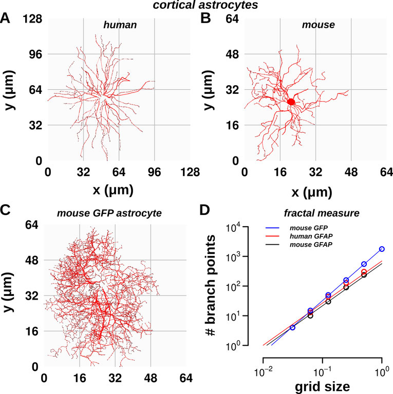
Figure 1: Computational methods for astrocyte morphology characterization. (A,B) Show tracings of GFAP-stained human and mouse astrocytes from the prefrontal cortex (Neuromorpho). (C) Shows the tracing of a transgenic GFP astrocyte from the mouse hippocampus (courtesy: Yukiko Goda, OIST). (D) Shows dimensional analysis with fractal box-counting. The red and black lines of human and mouse astrocytes have a slope of 1.42, indicating similar branch complexity. The blue line representing GFP astrocytes has a slope of 1.75 showing that GFP astrocytes have more labelled processes.
Data and code sharing: All the analyses performed for this project will be shared on our GitHub repository and in a Zenodo repository.
Since 2019, we have shared all our publication materials, including raw data and code, via Github (Dr. Garcia) and zenodo and figshare (both), and here we will take the same approach. We will use R, Python, and open-source libraries for analysis, incorporating tools developed by the Allen Institute via Jupyter Notebooks. We will use Binder to run the analysis directly from the GitHub repository without the need to install all the software dependencies, which will make the analysis easy to reproduce.
Replicability and reproducibility: Writing code in Python and R will make the analysis open and free, with detailed documentation for reusability and reproducibility. Moreover, using Jupyter Notebooks with Binder would enable analysis from the repository website avoiding the overhead of dependency installations.
FAIR standards: The four foundational principles of FAIR are Findability, Accessibility, Interoperability, and Reusability. We will incorporate these principles as we integrate our results with the original data.
We will share links to our repositories on Github and Zenodo in publications and websites to ensure findability. Zenodo's unique, persistent identifier will make it easy to find data. Writing our code in Jupyter Notebooks and using Binder would make our analysis easy to reproduce and replicate.
Our in-depth examination of human and mouse neocortices will yield three key findings that will significantly enhance our understanding of astrocytes in human brain function. First, it would test if astrocytes have a scalable architecture, indicating whether morphologies vary across regions or species while following consistent design rules. Second, it will reveal the local circuit factors associated with changes in astrocyte morphology. For instance, higher branching complexity might be associated with more astrocyte-synapse contacts. Third, these findings will test if a single theory of astrocyte scaling explains variations across species and regions, and how larger human astrocytes enhance function.
Our analysis will enhance understanding of astrocyte morphology, questioning the current view in neuroscience that astrocytes tile exclusive territories. The human H01 dataset shows that astrocytes do not consistently tile exclusive regions but instead show variable overlap across layers. By quantitatively describing astrocyte density and matching morphological variations to surrounding neuronal and vascular environments, we could test if astrocytes adapt to their environment. This will provide a model of how astrocytes, which might tile exclusively in the mouse cortex or in deeper layers, might not in humans or more superficial layers.
Understanding astrocyte architecture is crucial for comprehending brain health, both in normal conditions and in disorders like Alzheimer's and dementia. In most of these disorders, astrocyte morphologies are noticeably different in terms of branching complexity and size.
How does altered astrocyte morphology affect nearby synapses and neural function? Currently, the answer to this question remains challenging. There are no quantitative relationships describing the relationship between astrocytes and the local neuronal environment, which is a must if we are to develop theories that lead to a better understanding and therapies for those affected by neurological disorders.
Our project would take an important step in this path by providing vital quantitative information and theory of how astrocyte morphology is shaped to optimize function.
The team comprises Shyam Srinivasan Ph.D., a research scientist at the University of California San Diego, and a visiting scientist at Salk. Guadalupe C. Garcia Ph.D. is a postdoctoral fellow at the Salk and a visiting scholar at UC San Diego. Both share common interests in neuroanatomy, the morphology of astrocytes in particular, and in applying a theoretical approach.
Dr. Srinivasan began exploring how brains develop in his PhD in computer science and neuroscience at UC Irvine. In his postdoctoral work with Chuck Stevens he used neuroanatomy and theory to study scalable circuits. He is interested in how nature has designed neural circuits to be scalable at multiple levels and to function efficiently without wasting resources. He has a deep interest in how both constraints cooperate to optimize brains for each animal's environmental niche.
Dr. Garcia has a PhD in Biology, and a BS and MS in Physics. She has worked on several computational neuroscience projects generating and analyzing 3D EM reconstructions, characterizing mitochondria morphology and quantifying changes in presynaptic terminals after LTP. Since 2020, she has worked in Prof. Sejnowski's Lab, co-mentored by Prof. Kristen Harris, and our work will benefit from their combined expertise. Prof. Sejnowski is a pioneer in computational neuroscience and a recipient of multiple prestigious awards such as the Brain Prize.
