Peripheral nerve stimulation therapies deliver electrical signals to somatic nerves to restore limb function and to autonomic nerves for cardiovascular, cognitive, and inflammatory conditions, among others. However, it remains unclear how to define appropriate electrode geometries and stimulation parameters for effective therapy. We will reuse published nerve histology and in vivo data to parameterize and validate anatomically-realistic computational models of intrafascicular rat sciatic nerve stimulation. We will then use the validated models coupled with engineering optimization to design novel electrode geometries and stimulation parameters for selective nerve fiber activation. Selective stimulation will increase device efficacy and reduce side effects produced by stimulation of off-target nerve fibers. These complementary computational, anatomical, and in vivo studies provide the knowledge and technology that are required to advance neural prosthetics and bioelectronic medicines.
Aim 1: Implement and validate anatomically-realistic computational models of sciatic nerve stimulation
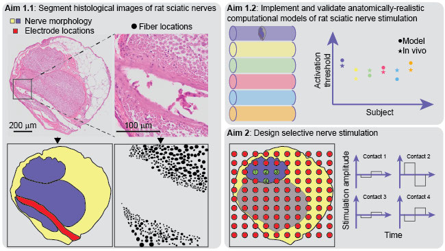
1.1: Neural anatomy
Data and tool reuse: We will analyze histological images of rat sciatic nerves from Open Science Framework: n=6 animals, OME-TIFF files, ~300 MB each. We will segment the images using the Segment Anything Model plugin to napari (Supp 1) and CellPose (Supp 2), which are freely-available.
Methods: For each histological image, we will segment the nerve morphology, the myelinated nerve fibers, and the location of each electrode contact.
Analyses: We will quantify the nerve morphology (nerve diameter, number of fascicles, fascicle diameters, perineurium thickness) and generate fiber diameter histograms.
Outcomes: The segmented images will serve as inputs to anatomically-realistic models in Aim 1.2.
1.2: Computational models of sciatic nerve stimulation
Data and tool reuse: We will use the open-source ASCENT and S-MF pipelines from GitHub (Supp 3 and 4) to model rat sciatic nerve stimulation. We will validate the modeled neural responses against in vivo activation thresholds (Supp 5) from Open Science Framework (~200 KB CSV file); these in vivo data and the histology in Aim 1.1 are for the same animals, which enables quantitative validation of model outputs.
Methods: We will use ASCENT to model intrafascicular rat sciatic nerve stimulation (n=6) using the segmented nerve morphologies from Aim 1.1, and we will use S-MF to simulate activation thresholds for the segmented fiber diameters and locations.
Analyses: We will compare modeled and in vivo activation thresholds. We will conduct sensitivity analyses of model parameters to identify values that generate the best accuracy for subject-specific models versus a generalized model.
Outcomes: The validated models of rat sciatic nerve stimulation will be used for device design in Aim 2.
Aim 2: Design selective nerve stimulation
Tool reuse: We will use the open-source “cajal” pipeline from GitHub (Supp 6) to optimize stimulation parameters for selective activation.
Methods: We will use the validated generalized model of rat sciatic nerve stimulation from Aim 1.2 with 10 x 10 electrode contacts over the nerve cross section. We will optimize the contact weightings and pulse width to activate selectively the nerve fibers in each fascicle (i.e., corresponding to a specific muscle group, e.g., the calf).
Analysis: Based on the optimized active contact configuration, we will simplify the electrode geometry and re-optimize the stimulation parameters.
Outcomes: The outcomes of this aim will be optimized electrode geometry and stimulation parameters for selective stimulation of each fascicle in the rat sciatic nerve.
Timeline
The available histology and in vivo data do not present constraints with respect to completeness or quality.
Dissemination
We will share the data on Open Science Framework (OSF) using the SPARC Data Structure (Bandrowski et al. 2021, doi:10.1101/2021.02.10.430563), with our image analysis methods on protocols.io. We will share the modeling tools on GitHub with documentation on Read the Docs. ASCENT includes a tool and documentation for generating model-based datasets that are compliant with the SPARC Data Structure (Supp 7). We will present the results at the Society for Neuroscience.
Outputs and file formats
Aim 1.1
Aim 1.2
Aim 2
FAIR principles and persistent identifiers
Findability: We will share our data, code, and models using established outlets. OSF and protocols.io generate a DOI upon publication, and the ASCENT GitHub is linked to Zenodo to generate version-specific DOIs.
Accessibility: Our shared data, code, and models will be freely and publicly available.
Interoperability: We will share the data and code in open and established file formats (OME-TIFF, CSV, TXT with Markdown, PY, JSON, DAT). ASCENT works on Windows, macOS, and Linux.
Reusability: Our datasets will include metadata, README’s, and protocols, as well as DOIs for provenance to the source histology and in vivo data. ASCENT is documented on Read the Docs.
Replicability and reproducibility
We will share our data on OSF under the CC-BY-4.0 license with complete documentation, and ASCENT is available under the GPLv2 license for non-commercial use. Thus, other scientists can either replicate the image analyses and models or directly use the shared segmentation masks and model outputs.
Peripheral nerve stimulation (PNS) therapies deliver electrical signals to the somatic system to restore limb function and to the autonomic system for cardiovascular, cognitive, inflammatory, metabolic, and painful conditions, among others. Since 1999, human trials of PNS have targeted somatic nerves to restore movement following spinal cord injury or sensation in persons with limb loss (Taghlabi et al. 2024, PMID:38237175).
FDA-approved PNS therapies use extraneural cuff electrodes. However, intrafascicular electrode arrays have long been used in preclinical and human studies, and the longevity and biocompatibility of intrafascicular devices have benefited from recent advances in materials and fabrication (e.g., Richie et al. 2024, PMID:38294928). Intrafascicular electrodes enable more selective activation of target fibers, require less energy to activate nerve fibers (thus lengthening battery life), and better enable scaling from preclinical to clinical studies by avoiding the increase in electrode-fiber distances (and thus activation thresholds) associated with cuff-based stimulation applied to larger nerves.
Multi-contact intrafascicular PNS devices require selection of many parameters to define appropriate electrode contact count, contact spacing, and stimulation parameters (i.e., “programming”). Programming poses a persistent challenge to effective treatment due to differences in effective stimulation parameters across patients and at different times for a given patient. Computational modeling and engineering optimization offer powerful approaches to design electrode geometry and stimulation parameters for selective nerve fiber activation to improve motor control and reduce side effects. However, quantitative validation of models of intrafascicular PNS is lacking.
The identified GREI dataset offers a robust and unique opportunity to advance model-based design of intrafascicular PNS: for each animal, it includes both in vivo recordings (activation thresholds and hind limb movements) and histological images. Thus, this dataset enables implementation of subject-specific anatomically-realistic models with experimental data for model validation, relating anatomy to neural and functional outcomes. We will conduct secondary analyses of these data (image segmentation), followed by data reuse to parameterize and validate a population of computational models.
These validated models will provide a strong foundation for further PNS development, including in vivo evaluation of the optimized PNS designs, comparisons of optimized selectivity with extraneural cuff electrodes vs. intrafascicular arrays, effects of intrafascicular electrode contact size on activation thresholds, and translation of PNS designs from preclinical experiments to clinical application.
Our team has extensive expertise and experience in the domains relevant to the proposed project, including neural engineering, peripheral nerve stimulation, multi-scale peripheral nerve anatomy, anatomically-realistic computational modeling, engineering optimization, in vivo electrophysiology, clinical translation, and statistics.
Dr. Frederick approached Dr. Pelot at the inaugural Gordon Research Seminar on Neuroelectric Interfaces after seeing her work on computational modeling of peripheral nerve stimulation. Dr. Frederick proposed reuse of her rigorous anatomy, electrophysiology, and functional data of rat sciatic nerve stimulation to parameterize and validate computational models to enable optimization.
Dr. Grill has conducted innovative high-impact translational research in neural stimulation for over 30 years, spanning computational modeling to preclinical in vivo experiments to translation clinical testing. Dr. Pelot has conducted neural engineering research for 12 years, specializing in computational modeling of peripheral nerve stimulation with complementary anatomical and in vivo studies. Mr. Marshall, Mr. Baumgart, Ms. Turk, and Mr. Hussain are developing state-of-the-art computational models of peripheral nerve stimulation to analyze mechanisms of action and develop novel therapies with improved clinical efficacy.
All team members have training and experience in statistical methods for experimental design and data analyses, as evidenced in their publications.
Three key considerations will ensure the project’s success: computational efficiency, model validation, and replicability.
Anatomically-realistic models are computationally demanding. However, (1) ASCENT standardizes and batches models of nerve stimulation, (2) S-MF is ~20,000x faster than the industry-standard model, (3) “cajal” optimizes parameters within minutes rather than days, and (4) our Duke Compute Cluster partition has a server-grade GPU, ~30k CPUs, and ~200 TB of RAM.
The utility of model-based design requires model accuracy. We previously quantitatively validated models rat, pig, and human vagus nerve stimulation (Supp 8, 9). We expect that our sensitivity analyses will successfully tune model parameters to produce quantitative match (<20% difference) between model and in vivo responses.
To ensure replicability of the model-based designs—in addition to data curation and sharing described in “Outcomes”—we will conduct multiple optimization runs with different random seeds.
