Cardiovascular diseases (CVDs) impact nearly half of American adults. Low-dose computed tomography (LDCT), effective for lung cancer screening, is underutilized for CVD risk analysis, despite being widely used since Medicare coverage began in 2015. Challenges in workflow and inconsistent diagnosis have hindered dual use for “radiation-free” CVD screening. This project aims to develop an AI model for CVD risk analysis using existing LDCT images, avoiding the need for costly, radiation-heavy cardiac scans. Utilizing data from GREI repositories, the model’s accuracy and reliability will be rigorously tested across diverse patient populations and institutions. Key innovations include turning LDCT into a dual-screening tool, creating an explainable AI model, and ensuring robust validation. This project promises significant advancements in early CVD detection for at-risk populations.
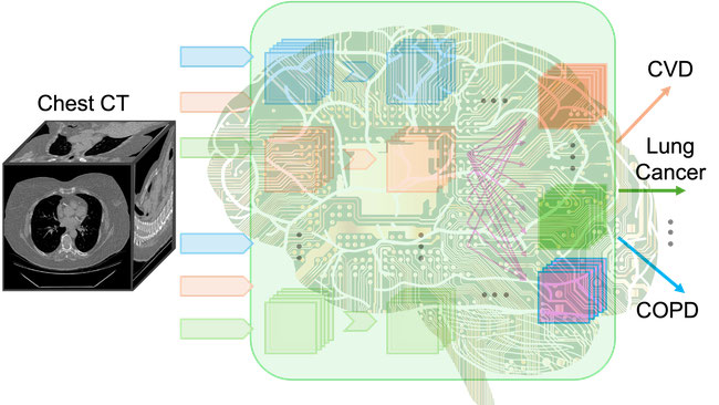
Several recent studies reported coronary artery calcification (CAC) grading in lung cancer screening scans is associated with mortality rate. In our preliminary study, we developed a deep learning CVD risk prediction model, trained with 30,286 LDCTs from the National Lung Cancer Screening Trial achieves promising performance on a separate test set of 2,085 subjects. By expanding the method to work on non-gated non-contrast chest CT, we will further increase the patient population that can benefit from the technology. The research aims of this project are as follows.
Aim 1: To build a large AI foundation model to harmonize imaging features. We will utilize the existing chest CT datasets, including but not limited to NLST, COPDGene, and RAD-ChestCT Dataset. We will reuse these large datasets to train the developed AI foundation model for CT image analysis.
Aim 2: CT radiomics-enhanced holistic CVD risk assessment. Several CVD risk scoring methods have been adopted in clinical practice, which rely on non-imaging information such as age, sex, race, medical conditions, and physiological measurements. When CAC is included, it needs to be measured from ECG-gated cardiac CT. Without specific imaging markers, those methods focus more on population statistics than personalized risk prediction for individual subjects. Enhancing the existing clinical tools with image-based assessment will utilize both imaging and non-imaging markers to provide a holistic prediction.
Aim 3: Validation of accuracy, reliability, and generalizability. The validation will be performed in patients with LDCT for lung cancer screening from GERI repositories. We will use patient record and follow up results as the labels for evaluation.
Upon successful completion, the project will provide critical, yet “radiation-free” tools for CVD risk assessment of high-risk subjects. It will enhance the knowledge about the value of LDCT for CVD risk estimation, overcome the limitations of the currently applied analysis methods, and advance chest CT-related healthcare. The innovative deep learning tools and training strategies can be extended to evaluate risks of related diseases and other AI-based clinical decision support systems.
1. Research Findings and Summary of Expected Outcomes:
- The project aims to develop an advanced deep learning foundation model to analyze cardiovascular disease (CVD) risks from low-dose computed tomography (LDCT) scans originally intended for lung cancer screening. The focus is on creating an explainable and trustworthy model that integrates both imaging and non-imaging factors to provide a more comprehensive CVD risk assessment.
- The expected outcome is a tool that can enable "radiation-free" CVD screening as a secondary benefit of existing lung cancer LDCT scans, thus reducing the need for additional scans and radiation exposure. This project aims to improve early CVD detection, potentially preventing severe cardiovascular events in high-risk individuals.
2. How Findings Will Be Shared and Locations:
- The research findings will be disseminated through publications in peer-reviewed journals and presentations at conferences related to medical imaging, cardiovascular research, and AI in healthcare.
- To facilitate further research and adoption, the project will release its source code and developed tools on platforms like GitHub. This will include tools for implementing the foundation model for CVD risk estimation, which can be used by other researchers and clinicians.
- The project also plans to develop an online application, making it accessible to researchers with clinical backgrounds who may not have extensive computational resources. This app will allow for easy deployment and integration of the research findings into clinical practice.
3. Addressing FAIR and CARE Principles:
- FAIR Principles: The project commits to making the data and tools Findable, Accessible, Interoperable, and Reusable by sharing source codes, data models, and documentation. This will include using open-access repositories like GitHub and providing detailed metadata to ensure easy reuse and integration.
- CARE Principles: Where relevant, the project will ensure that data sharing respects the principles of Collective benefit, Authority to control, Responsibility, and Ethics, especially concerning the use of patient data. Privacy and ethical considerations will be prioritized in the dissemination of clinical data.
4. Addressing Replicability and Reproducibility:
- The project emphasizes the reproducibility of its foundation model through systematic testing and validation across different clinical sites, patient demographics, and scanner technologies. By using diverse datasets and validating models in multiple settings, the research aims to ensure that results are not only replicable but also generalizable.
- To aid in replication, the project will share detailed protocols, annotated datasets, and training procedures, allowing other researchers to reproduce the methods and verify results. This openness is intended to build trust in the developed tools and promote widespread use.
The research aims to advance the use of low-dose computed tomography (LDCT) scans as a dual-purpose tool for both lung cancer and cardiovascular disease (CVD) risk assessment. By leveraging deep learning foundation models, the project contributes to the integration of medical imaging with AI, a cutting-edge area in biomedical engineering. It enhances the ability to extract meaningful clinical insights from routine imaging, which could serve as a model for other cross-discipline applications of AI in medical diagnostics.
The project could significantly improve the diagnostic capabilities for identifying high-risk CVD patients without needing additional, dedicated cardiac imaging. By utilizing LDCT scans that patients are already undergoing for lung cancer screening, the approach enables a more comprehensive assessment of both lung and cardiovascular health from a single scan, thereby improving early detection of at-risk patients.
The integration of CVD risk analysis into routine lung cancer screening has the potential to serve as a preventive measure for a large segment of the population. Patients identified as high-risk could receive recommendations for lifestyle changes, such as smoking cessation, diet improvements, or increased physical activity, aimed at reducing their overall cardiovascular risk. This preventive approach aligns with public health goals of reducing the incidence of heart disease and related complications.
Overall, the research project holds the potential to make substantial contributions to both scientific understanding and practical clinical applications, making it a pivotal effort in enhancing the intersection of cancer screening and cardiovascular care. It emphasizes the role of interdisciplinary innovation in solving complex health challenges.
The proposed project is interdisciplinary and translational, which combines the synergistic research strengths of the collaborative team. Dr. Pingkun Yan (RPI) is an expert in machine learning and medical image computing with both academic research and industrial R&D experience; Dr. Mannudeep K. Kalra (MGH, Harvard) is a radiologist specializing in cardiac and thoracic imaging with a focus on lung cancer screening (radiation dosimetry, protocol optimization, clinical workflow, and CAD); Dr. Ge Wang (RPI) is a leading CT imaging expert dedicated to deep tomographic reconstruction and radiomic analysis; Dr. Xuanang Xu (RPI) is an image analysis expert with strong skills in machine learning and artificial intelligence. Over the past several years, the team has worked together closely and effectively with a number of joint publications on LDCT image analysis and denoising. In this project, we will develop advanced medical image analysis methods for LDCT-based CVD risk quantification with the goal of eventually translating to clinical use.
The project benefits from a strong multidisciplinary team, including experts in AI, medical imaging, and radiology, enabling comprehensive problem-solving and iterative model improvements. Furthermore, by adopting an open-source approach, the project will share its code and tools, fostering broader collaboration and enabling the scientific community to validate and build upon its findings. This transparency ensures reproducibility and enhances the potential impact of the research. The project also leverages the powerful computing equipment at RPI and the research center co-directed by Drs. Yan and Wang.
